GLAUCOMA
 Glaucoma is a condition that causes damage to your eye’s optic nerve and gets worse over time. It’s often associated with a buildup of pressure inside the eye.
The increased pressure, called intraocular pressure, can damage the optic nerve, which transmits images to the brain. If damage to the optic nerve from high eye pressure continues, glaucoma will cause permanent loss of vision.
Glaucoma usually occurs when pressure in your eye increases. This can happen when eye fluid isn’t circulating normally in the front part of the eye. Normally, this fluid, called aqueous humor, flows out of the eye through a mesh-like channel. If this channel becomes blocked, fluid builds up, causing glaucoma.
Less common causes of glaucoma include an injury to the eye, a severe eye infection, blockage of blood vessels in the eye, inflammatory conditions of the eye, and occasionally eye surgery to correct another condition
SYMPTOMS OF GLAUCOMA
In the beginning, open-angle glaucoma has no symptoms and it causes no pain. Vision stays normal. Glaucoma can develop in one or both eyes.
Without proper glaucoma treatment, people with this disease will slowly lose their peripheral vision. As glaucoma remains untreated, people may miss objects to the side and out of the corner of their eye. It is like looking through a tunnel. Over time, glaucoma steals the straight-ahead central vision until no vision remains.
RISK FACTORS FOR GLAUCOMA
Anyone can develop glaucoma. Some people, listed below, are at higher risk than others:
- African Americans over age 40
- Everyone over age 60, especially Mexican Americans
- People with a family history of glaucoma
A comprehensive dilated eye exam can reveal more risk factors, such as high eye pressure, thinness of the cornea, and abnormal optic nerve anatomy. In some people with certain combinations of these high-risk factors, medicines in the form of eye drops reduce the risk of developing glaucoma by about half.
HOW IS GLAUCOMA DETECTED?
Glaucoma is detected through a comprehensive dilated eye exam that includes the following:
- Visual acuity test. This eye chart test measures how well you see at various distances.
- Visual field test. This test measures your peripheral (side vision). It helps your eye care professional tell if you have lost peripheral vision, a sign of glaucoma.
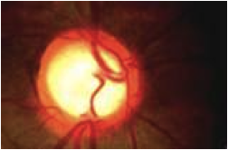
- Dilated eye exam. In this exam, drops are placed in your eyes to widen, or dilate, the pupils. Your eye care professional uses a special magnifying lens to examine your retina and optic nerve for signs of damage and other eye problems. After the exam, your close-up vision may remain blurred for several hours.
- Tonometry is the measurement of pressure inside the eye by using an instrument called a tonometer. Numbing drops may be applied to your eye for this test. A tonometer measures pressure inside the eye to detect glaucoma.
Pachymetry is the measurement of the thickness of your cornea. Your eye care professional applies a numbing drop to your eye and uses an ultrasonic wave instrument to measure the thickness of your cornea.
TREATMENT
Glaucoma treatments include eye drops, laser trabeculoplasty, conventional surgery, or a combination of any of these. While these treatments may save remaining vision, they do not improve sight already lost from glaucoma. There is no cure for glaucoma. The vision lost from glaucoma disease cannot be restored.
EYE DROPS FOR GLAUCOMA.
The eye drops are prescription medicines. These either reduce the formation of fluid in the front of the eye or increase its outflow.
LASER SURGERY FOR GLAUCOMA.
Laser surgery for glaucoma slightly increases the outflow of the fluid from the eye in open-angle glaucoma or eliminates fluid blockage in angle-closure glaucoma.
Types Of Laser Surgery For Glaucoma:
- Trabeculoplasty – a laser is used to pull open the trabecular meshwork drainage area. An iridotomy is performed, where a tiny hole is made in the iris, allowing the fluid to flow more freely.
- Cyclophotocoagulation – a laser beam treats areas of the middle layer of the eye, reducing the production of fluid.
- Microsurgery for glaucoma. In an operation called a trabeculectomy, a new channel is created to drain the fluid, thereby reducing intraocular pressure that causes glaucoma.
At this time, loss of vision caused by glaucoma is irreversible and cannot be restored. However, successfully lowering eye pressure can help prevent further visual loss from glaucoma. Most people with glaucoma do not go blind if they follow their treatment plan and have regular eye exams.